
As part of my fourth year, I chose to do an elective in forensic pathology at the medical examiner’s office. Many people, including my peers, wondered why a bubbly, optimistic future obstetrician-gynecologist would choose to see death on a daily basis. I, on the other hand, had been waiting for this rotation since my first year. And it was medicine like I had never seen before.
 On my first day, I was given a detailed schedule, and each day I worked with a different pathologist down in the basement. Currently, there are 14 forensic pathologists working at the medical examiner’s (ME) office! I was told there is room for up to 17 physicians because of the vastness of Maricopa County. Maricopa is the 4th largest county in the nation, spanning in over 9200 square miles. Each day, 3-4 physicians are assigned to work on autopsy cases while the others work on paperwork for their cases. Due to the sheer size of the county, there are a multitude of cases that come in. On top of this, some cases are termed “expedites,” which include babies, death by water submersion, and family emergencies requiring expedition. So in addition to the four cases assigned to the physicians doing cases that day, they must also pick up the expedites, leading to a very busy day. I arrived at 7 am for a daily meeting with the physicians, lead forensic tech, death investigators, and forensic photography. We reviewed photographs of death scenes for each case before heading downstairs. As part of my rotation, I kept my own case journal of my cases each day and wrote down salient points from the autopsy.
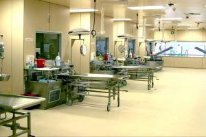
I truly appreciated the role of personal protective equipment (PPE) on this rotation. Putting on PPE was a morning ritual that took at least 5-7 minutes for new people (like me). Every morning, I plunged my legs and feet into full length booties, wore an XXL scrub gown along with plastic white sleeves. I then placed an extra apron on top of my scrubs (these would be changed between every case), put on a face shield and scrub cap, and donned an N95 mask with two layers of gloves. As I walked into the examination rooms each morning, the forensic techs would already have the first case unzipped from the body bag and ready to be examined. We first began with an external examination, noting any defects on the skin, looking at tattoos to confirm identity, and documenting medical interventions. At this time, the techs would also obtain samples of vitreous fluid from the eye for postmortem electrolyte and glucose analysis. I helped the techs transfer the body to the table. Then, the autopsy began with a traditional Y-shaped incision from sternum to pubis. Every organ was weighed and sliced by the pathologist, and samples would be set aside in jars of formalin for histology. Once the pathologist began examining organs, the techs would move on to opening the skull with the bone saw.
There was a huge aspect of mystery to a majority of our cases. Decedents would be found at home or outside, and their immediate cause of death was unknown. Several of these cases ended up having > 70% stenosis of the left anterior descending artery and “ASCVD” would be the likely cause of death. Other cases were full of surprises. In one elderly male, we discovered two massively dilated ureters and renal calyces, only to find that he had an enormous tumor in his bladder that was invading into the pelvis. In another case, the patient was found dead in their home, and upon cutting into the brain, we saw a glaring hemorrhagic stroke in the left hemisphere. I felt as if I were seeing things from my first- and second-year lectures that I never got to connect to a clinical scenario. I saw more “trauma” than I did on my trauma surgery rotation. We had multiple decedents who were part of a motor vehicle accidents. In one case, I was able to run my hand over a complete spinal cord transection and palpate an atlanto-odontoid separation as well as a cervico-medullary tear in the brainstem. The pathologists did a wonderful job of including me in cases that were unique. I removed a bullet fragment from the back of a skull, felt the auto-infarcted spleen of a decedent who had sickle cell disease, and saw my first horseshoe kidney.
During one of my weeks, I spent time with the death investigations team and went to various death scenes all over Maricopa County. The death investigations team is composed of extremely hardworking individuals who endure extreme heat, putrid smells, and filthy environments all in a day’s work—and with far less PPE than I was used to. Our scenes included homicides with the CSI unit, sudden infant deaths, suicides, decomposing bodies, and more. We went to anything that was under the umbrella of needing an autopsy at the ME office. Our investigations included photography, placement of the decedent in the body bag, and discussion with nearby family and friends to understand the details of the death. This also involved counseling grieving family members.
On my last week of the rotation, I spent a day with the forensic odontologist and the forensic anthropologist (the real life Bones!). Dr. Piakis, the in house forensic odontologist, walked me through how he compares dental imaging of the deceased to previous dental records. In this way, he confirms identification. Dr. Fulginiti, the forensic anthropologist, led me up to her lab where her interns were taking a skull out of a Crock-Pot that had been sitting in solution to erode the tissue from the bones. They were subsequently going to analyze the potential cause of blunt force trauma to the skull. She laid out an entire “bone workshop” for me, with different stations of bones from different blunt force trauma injuries. I saw a chainsaw injury to the clavicle, gunshot wounds to the skull, penetrating trauma to the skull, and more.
This rotation was fascinating and challenging in more ways than one. Some days included emotionally heavy cases that especially pulled at my heartstrings: sudden infant death, infant or pediatric trauma, teenage suicides. The forensics team always showed respect and worked meticulously to perform thorough autopsies for their cases. After doing multiple capstones at the ME office during my first year, I knew what I was getting myself into. I came in hoping to see interesting cases and learn about death-scene investigations. Upon leaving the rotation, I felt that I gained an ability to see death in a new light. I also feel more equipped to face loss in my future field of obstetrics and gynecology. Forensic pathology is truly a unique field, and I recommend students try out this elective during fourth year!
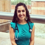
Nura Sobhanian is a medical student at The University of Arizona College of Medicine – Phoenix, Class of 2018. She is originally from San Clemente, California, and is currently interested in pursuing a career in OB/Gyn. She likes to rock a pair of Rainbow sandals in her free time, and you can find her with a book at any local coffee shop in metro Phoenix.