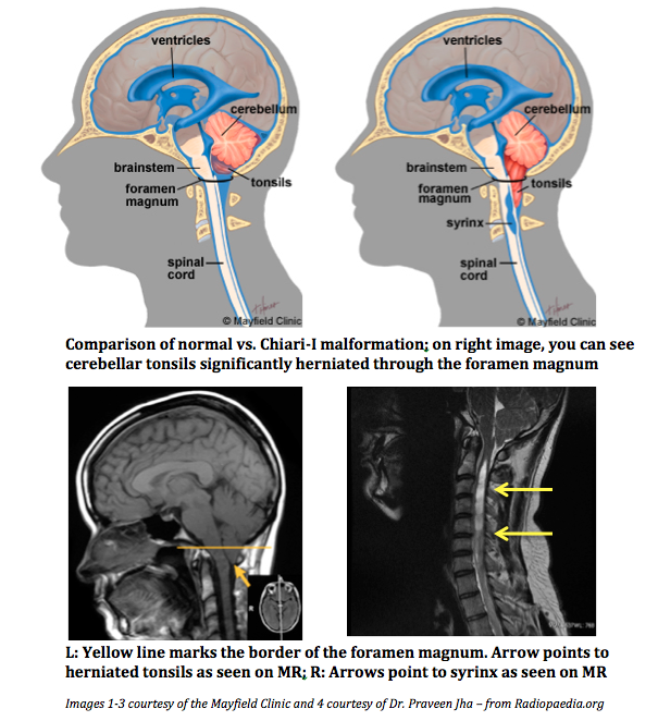
 Arnold-Chiari malformations describe a cluster of congenital neurologic defects characterized by the downward displacement of cerebellar tonsils through the foramen magnum. Tonsillar herniation can lead to serious neurologic sequelae by obstruction of cerebrospinal fluid (CSF). In Chiari malformation type-I (CM-I), the typical course is surprisingly benign—patients typically are asymptomatic, and the Chiari malformation is discovered incidentally on imaging [1-2]. CM-I is diagnosed if the cerebellar tonsils are displaced ≥ 5 mm below the foramen magnum. Frequently, it presents with a syrinx, a cyst in the spinal canal formed from impaired flow and a subsequent altered pressure differential of CSF due to this obstruction. Thus, asymptomatic CM-I patients can be managed conservatively with careful surveillance for new-onset symptoms [3-4].
If CM-I patients do become symptomatic, they may present with occipital headaches, nuchal pain, ataxia, weakness, dysphagia, numbness/tingling, and drop attacks [1]. These symptoms can be incapacitating, preventing patients from attending to school/work responsibilities and enjoying daily activities. Symptom presentation with altered CSF dynamics (usually in the form of a syrinx) is a clear indication for neurosurgical intervention [5]. A suboccipital craniectomy with C1 laminectomy is performed to relieve the pressure on the cerebellar tonsils, thereby restoring normal CSF flow. However, it is controversial whether surgery should be considered at an earlier stage before symptom presentation or imaging changes. Neurologic symptoms are irreversible, further underscoring the importance of timely intervention for optimal outcomes and quality of life.
My scholarly project (SP) aims to explore the use of imaging changes as a potential predictive indicator in surgical outcomes of symptomatic CM-I patients who have undergone the suboccipital craniectomy. I am working with Dr. Jorge Arango at the Barrow Neurological Institute (BNI) at Phoenix Children’s Hospital. By analyzing a retrospective cohort, I hope to find key correlations between MR imaging changes (magnitude of tonsillar descent, syrinx, third ventricular space, skull base angle, ventral canal encroachment) and surgical outcomes as determined by post-op progress notes and the Chicago Chiari Outcome Scale (CCOS). The CCOS is a validated scale to evaluate CM-I patients post-op in four categories: pain symptoms, non-pain symptoms, functionality, and surgical complications. Higher composite scores indicate better overall outcomes [6]. Successful completion of this project will hopefully provide valuable evidence for the use of imaging changes in determining the most efficacious period for surgery. Furthermore, timely intervention may result in a more dramatic resolution of debilitating symptoms and uncomplicated recovery, leading to a quick return to day-to-day activities and significantly improved quality of life.
My interest in neuroscience was sparked during my undergraduate time at UCSD. I had an opportunity to learn from amazing professors like Dr. Ramachandran (Google: phantom limbs) in classes focused entirely on brain damage, cognitive neuroscience, and even the frontal lobe and cognitive control. Our brain is a fragile organ that composes everything we are as biologic entities and as people. I saw firsthand how dysfunction to even a small portion of the brain could adversely change a patient’s life. I hope my project can contribute to the growing knowledge of a burgeoning neuroscience field, ultimately leading to better ways to take care of our patients.
- Aitken LA, Lindan CE, Sidney S, et al. Chiari type I malformation in a pediatric population. Pediatr Neurol. 2009;40(6):449-454.
- Benglis D, Jr, Covington D, Bhatia R, et al. Outcomes in pediatric patients with Chiari malformation type I followed up without surgery. J Neurosurg Pediatr. 2011;7(4):375-379.
- Nishizawa S, Yokoyama T, Yokota N, Tokuyama T, Ohta S. Incidentally identified syringomyelia associated with chiari I malformations: Is early interventional surgery necessary? Neurosurgery. 2001;49(3):637-40; discussion 640-641.
- Novegno F, Caldarelli M, Massa A, et al. The natural history of the Chiari type I anomaly. J Neurosurg Pediatr. 2008;2(3):179-187.
- Schijman E, Steinbok P. International survey on the management of Chiari I malformation and syringomyelia. Childs Nerv Syst. 2004;20(5):341-348.
- Aliaga L, Hekman KE, Yassari R, et al. A novel scoring system for assessing Chiari malformation type I treatment outcomes. Neurosurgery. 2012;70(3):656-664; discussion 664-665.

Dan Kim is a second-year medical student at The University of Arizona College of Medicine – Phoenix. He completed his BA in psychology at UC San Diego, realizing halfway through that he had the masochistic desire to attend medical school. Dan is passionate about the use of mobile technology in healthcare, clinical informatics, the history of medicine, and medical education. In the future, Dan hopes to pursue an academic career that will allow him to integrate his passion for research and teaching while taking good care of his patients. Contact him at dankim[at]email.arizona.edu.