The use of modern photography in medicine was embraced soon after its birth by Louis Daguerre in 1839. In the very same year, French bacteriologist and doctor Alfred Francois Donne photographed leukemia under a microscope. The Scottish painter David Octavius Hill produced a portrait in 1847 depicting a woman with a large goiter.1,2 Medical photography allows an objective visual portrayal of the patient’s condition, which led many medical specialties to quickly integrate photography into the way medicine is practiced and documented.
At a similar time in history, physicians and scientists were trying to solve a tangential problem. In the early 1800s, no reliable methods existed to peer inside body cavities without inflicting damage. The gastrointestinal (and urogenital) tract is not straight, and it’s dark in there. It wasn’t until 1806 when Dr. Philipp Bozzini of Germany assembled his Lichtleiter (German for light conductor). The Bozzini Endoscope, as it is also known, was the first device used to inspect the interior of the human body with an internal light source. Originally designed to examine the larynx, and later adapted for urological cases, this device used a candle and mirrors to allow doctors to view body cavities.3 All of this was accomplished before the invention of the light bulb!
In 1853, Dr. Antonin Jean Desormeaux went on to improve Bozzini’s design by using gas lamps instead of candles to perform minor procedures through endoscopes. These devices further transformed, when Dr. Hans Christian Jacobaeus of Sweden performed the first laparoscopic procedure on humans in 1910.4 Now, physicians could peer inside the body through “man-made” holes in the abdomen.
Following laparoscopy, the term arthroscopy was coined when Dr. Severin Nordentoft presented his version of an endoscope used to examine the interior of the knee in 1912.5 The early 1900s launched the beginning of the “Incandescent era” of endoscopy, where gas lamps were replaced by light bulbs and mirrors gave way to lenses. It was in the 1950s, however, that fiber-optic technology slowly ushered in a new era of endoscopy.
Before its use in transatlantic telecommunications, fiber-optics were used in endoscopy. Fiber optics took advantage of the physical concept of total internal reflection with the use of materials such as pure glass or plastic to transmit photons across cables. It was in 1958 that engineer Larry Curtiss and Dr. Basil Hirschowitz developed the flexible fiber-optic endoscope.6 This device pioneered the field of endoscopy by its reduced size, improved field of vision, and enhanced functionality, all of which contributed to lowering patient discomfort while maximizing image quality.
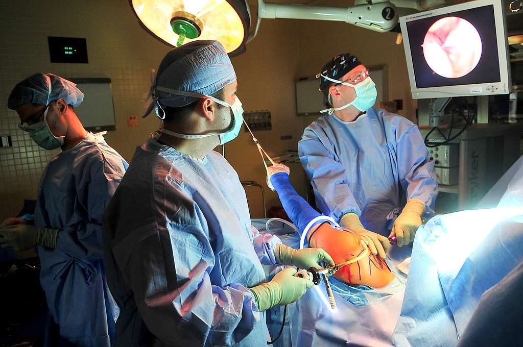
The “fiber-optic era” of endoscopy transformed once more with the invention of the charge-coupled device (CCD), where photons were converted into electrical charges with the use of a sensor array and linked capacitors. Converting the image from fiber-optic to electronic allowed endoscopy to become a group activity, since the image could then be displayed on a television screen for all to see.6 Following this, endoscopy entered into its golden era, and some medical fields like gastroenterology were forever changed.
Endoscopy and its technology continuously evolve year by year. “Chip-on-tip” sensors are now as small as 1mm in diameter, surgeries are being performed with robotic assistance, and some high-definition endoscopes can produce images with resolutions of up to 1 million pixels.7 It is important to note, however, that this technology is not without associated risks. Some endoscopic procedures draw massive amounts of power for the fiber-optic light source and can cause fires and burns. Malfunctions can cause the tip to reach temperatures of over 500 degrees Fahrenheit.8,9 In addition, the cables associated with endoscopes can put OR staff at risk of falls and cause sterility concerns. To combat this, some companies like Lazurite have developed a wireless arthroscopic camera system to declutter the surgical field and relief stress on OR staff.10
What if a patient cannot tolerate, or refuses, an endoscopic procedure? After its FDA approval in 2004, “Capsule Endoscopy” began to increase in prevalence among gastroenterologists.11 In a capsule endoscopy, a small pill-shaped wireless camera is swallowed. The camera then captures and transmits images as it passes through the GI tract for the doctor to later evaluate. There are many advantages to this approach, with the most important being the lack of procedural sedation. In addition, the newer dual camera systems can shoot up to 36 frames per second, involve a 340 degree field of view, and can use steerable capsules/magnets for navigation.11
Capsule endoscopy and its tech have evolved to visualize every section of the GI tract, but there are significant challenges and limitations with this technology. Transmit times in the GI tract are fast in places like the esophagus, and slow in the colon. Currently, there is no method of tissue sampling with capsule endoscopy. Despite these limitations, however, this technology has promise. Some experts envision a future where colonoscopies will become a procedure that is only recommended for patients that require a biopsy, reducing medical waste and cost for the patient.
In less than two hundred years, medical photography and endoscopy have transformed the way doctors learn and practice medicine. Although it is impossible to predict the future of endoscopy 50 years from now, it is safe to assume that interventions will continue to become less invasive and more comfortable for the patient.
References:
1. Thorburn AL. Alfred François Donné, 1801-1878, discoverer of Trichomonas vaginalis and of leukaemia. Br J Vener Dis. 1974;50(5):377-380. doi:10.1136/sti.50.5.377
2. A Brief History of Early Medical Photography – Clinical Correlations. Accessed June 27, 2023. https://www.clinicalcorrelations.org/2016/09/30/a-brief-history-of-early-medical-photography/
3. SPANER SJ, WARNOCK GL. A Brief History of Endoscopy, Laparoscopy, and Laparoscopic Surgery. https://home.liebertpub.com/lap. doi:10.1089/lap.1997.7.369
4. Hatzinger M, Kwon ST, Langbein S, Kamp S, Häcker A, Alken P. Hans Christian Jacobaeus: Inventor of human laparoscopy and thoracoscopy. J Endourol. 2006;20(11):848-850. doi:10.1089/end.2006.20.848
5. de Mello Granata GS. A hundred years of knee arthroscopy. Rev Bras Ortop. 2015;47(6):684. doi:10.1016/S2255-4971(15)30022-7
6. Sivak MV. Gastrointestinal endoscopy: past and future. Gut. 2006;55(8):1061-1064. doi:10.1136/gut.2005.086371
7. Jang JY. The Past, Present, and Future of Image-Enhanced Endoscopy. Clin Endosc. 2015;48(6):466-475. doi:10.5946/ce.2015.48.6.466
8. Quick Safety Issue 69: Preventing light source-related burns from laparoscopy, thoracoscopy and arthroscopy | The Joint Commission. Accessed June 28, 2023. https://www.jointcommission.org/resources/news-and-multimedia/newsletters/newsletters/quick-safety/quick-safety-issue-69/#.ZDVwcvbMIhd
9. Chitnavis J. Silent burn: the hidden danger and effects of bright light from fibre-optic cables in arthroscopic knee surgery. J Surg Case Rep. 2020;2020(4):rjaa068. doi:10.1093/jscr/rjaa068
10. Williams J, Ustin J, Calcei J, Gardiner K, Voos J, Polito P. Wireless vs. Wired Arthroscopic Cameras: A Comparative OR Simulation Study of Efficiency, Cost Per Case, Setup, and Takedown. J Orthopaedic Experience & Innovation. Published online December 12, 2022. Accessed June 28, 2023. https://journaloei.scholasticahq.com/article/38662-wireless-vs-wired-arthroscopic-cameras-a-comparative-or-simulation-study-of-efficiency-cost-per-case-setup-and-takedown
11. Oka P, McAlindon M, Sidhu R. Capsule endoscopy – a non-invasive modality to investigate the GI tract: out with the old and in with the new? Expert Review of Gastroenterology & Hepatology. 2022;16(7):591-599. doi:10.1080/17474124.2022.2089113

Benson Lagusis is a member of the University of Arizona College of Medicine - Phoenix Class of 2026. He graduated with a B.S. in Exercise Science from Northern Arizona University before earning a Master's degree in Biomedical Sciences from Midwestern University. Benson played baseball in college, and is interested in sports medicine and health promotion/education. During his free time, Benson is an avid hiker, reader, and Netflix binge-watcher.